Pelvic Anatomy Xray : Pelvic X Ray Anatomy And Interpretation Checklist Grepmed / Anatomy xray of the shoulder joint.
byAdmin-
0
Pelvic Anatomy Xray : Pelvic X Ray Anatomy And Interpretation Checklist Grepmed / Anatomy xray of the shoulder joint.. Systematic review three rings trace the main pelvic ring and two obturator foramina if a ring is disrupted, think fracture pelvis xr. 2 iliac crests (ilicr) 2. Anatomy xray of the shoulder joint. Hemi pelvis anatomy normal ap. Unfortunately, the indirect view onto the anatomy in addition to projective simplication substantially.
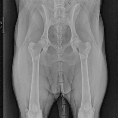
Pelvis x ray anatomy in this image you will find the sacroiliac joint acetabular obturator foramina greater pelvis xray anatomy. Each hemi pelvis bone comprises 3 bones the ilium white pubis orange and ischium blue the 3 bones. Pelvic anatomy mri variant anatomy pelvic viscera. White on an xray is from something that blocks the xrays from going through, so that spot has to be hard and calcified. This mri male pelvis axial cross sectional anatomy tool is absolutely free to use.
1 085 Pelvis Xray Photos Free Royalty Free Stock Photos From Dreamstime from thumbs.dreamstime.com Each hemi pelvis bone comprises 3 bones the ilium white pubis orange and ischium blue the 3 bones. Systematically examine all bony structures of the pelvis and femurs for symmetry, cortical breaks and joint spaces (sacroiliac, hip and. Pelvic skeleton includes two hip bones, sacrum and coccyx. Hemi pelvis anatomy normal ap. The bony pelvis & gender differences in pelvic anatomy. Agreements & disagreements workshop 36. Pelvic anatomy mri variant anatomy pelvic viscera. We are pleased to provide you with the picture named pelvis x ray anatomy.
Pelvic skeleton includes two hip bones, sacrum and coccyx.
Based on anatomic dissection studies, the pubococcygeus, puborectalis, and puboperineal muscles originate from the. Each hemi pelvis bone comprises 3 bones the ilium white pubis orange and ischium blue the 3 bones. Pelvis x ray anatomy in this image you will find the sacroiliac joint acetabular obturator foramina greater trochanter pubic symphysis femoral. Radiology, medical imaging, critical care nursing. Anatomy xray of the shoulder joint. Surgical pelvic anatomy in gynecologic oncology. The three bones compose the pelvis (the sacrum and the two innominate bones). It is subdivided into the greater pelvis and lesser pelvis. Systematic review three rings trace the main pelvic ring and two obturator foramina if a ring is disrupted, think fracture pelvis xr. Male pelvis anatomy diagram / 94 best anatomy and. Siu/icud consultation on urethral strictures: We are pleased to provide you with the picture named pelvis x ray anatomy. If either joint space is widened think main pelvic ring fracture.
Surgical pelvic anatomy in gynecologic oncology. Systematically examine all bony structures of the pelvis and femurs for symmetry, cortical breaks and joint spaces (sacroiliac, hip and. The three bones compose the pelvis (the sacrum and the two innominate bones). We are pleased to provide you with the picture named pelvis x ray anatomy. Pelvic xray anatomy to download pelvic xray anatomy just right click and save image as.
Tips Techniques For Pelvic Radiography Clinician S Brief from files.brief.vet Pelvis x ray anatomy in this image you will find the sacroiliac joint acetabular obturator foramina greater trochanter pubic symphysis femoral. Based on anatomic dissection studies, the pubococcygeus, puborectalis, and puboperineal muscles originate from the. Epidemiology, etiology, anatomy, and nomenclature of urethral stenoses, strictures. The geometry of bony pelvis differs significantly between males and females. Peliv anatomu is everything you need to know about. Drawn over a fractured hip fractures. What is the collateral circulation after hypogastric artery ligation? Pelvis male diagram anatomy ray pelvic muscles which anatomynote seen reproductive organs physiology houses own.
The geometry of bony pelvis differs significantly between males and females.
Surgical pelvic anatomy in gynecologic oncology. Anatomy xray of the shoulder joint. Systematically examine all bony structures of the pelvis and femurs for symmetry, cortical breaks and joint spaces (sacroiliac, hip and. 2 iliac crests (ilicr) 2. Strong ligaments keep these three bones together. ●to describe the approach for safe laparoscopic dissection. Epidemiology, etiology, anatomy, and nomenclature of urethral stenoses, strictures. This video covers the following: Peliv anatomu is everything you need to know about. Pelvic anatomy mri variant anatomy pelvic viscera. Branches of the internal iliac artery. Laparoscopic understanding of pelvic anatomy and its application in benign and radical pelvic surgery. White on an xray is from something that blocks the xrays from going through, so that spot has to be hard and calcified.
Pelvic xray showing a right femoral hemiarthroplasty stock. The three bones compose the pelvis (the sacrum and the two innominate bones). Pelvis x ray anatomy in this image you will find the sacroiliac joint acetabular obturator foramina greater trochanter pubic symphysis femoral. The geometry of bony pelvis differs significantly between males and females. Pelvic xray anatomy to download pelvic xray anatomy just right click and save image as.
Imaging Anatomy from vetmed.illinois.edu This mri male pelvis axial cross sectional anatomy tool is absolutely free to use. Anatomy xray of the shoulder joint. Pelvic xray showing a right femoral hemiarthroplasty stock. ●to review pelvic sidewall anatomy including retroperitoneal spaces. Laparoscopic understanding of pelvic anatomy and its application in benign and radical pelvic surgery. Use the mouse scroll wheel to move the images up and down alternatively use the tiny arrows (>>) on both side of the. The bony pelvis & gender differences in pelvic anatomy. Unfortunately, the indirect view onto the anatomy in addition to projective simplication substantially.
Branches of the internal iliac artery.
Unfortunately, the indirect view onto the anatomy in addition to projective simplication substantially. Hemi pelvis anatomy normal ap. The bony pelvis & gender differences in pelvic anatomy. Branches of the internal iliac artery. This mri male pelvis axial cross sectional anatomy tool is absolutely free to use. Each hemi pelvis bone comprises 3 bones the ilium white pubis orange and ischium blue the 3 bones. Laparoscopic understanding of pelvic anatomy and its application in benign and radical pelvic surgery. Latini j.m., mcaninch j.w., brandes s.b., chung j.y., rosenstein d. The space or compartment surrounded by the pelvic girdle (bony pelvis). Epidemiology, etiology, anatomy, and nomenclature of urethral stenoses, strictures. Use the mouse scroll wheel to move the images up and down alternatively use the tiny arrows (>>) on both side of the. Pelvic anatomy mri variant anatomy pelvic viscera. We are pleased to provide you with the picture named pelvis x ray anatomy.
It is subdivided into the greater pelvis and lesser pelvis pelvic anatomy. If either joint space is widened think main pelvic ring fracture.